
Rheumatic heart valve disease is chronic damage to one or more heart valves following rheumatic fever — an immune reaction to a streptococcal throat infection that was not properly treated. Its most distinctive feature is the delay: the damage does not appear during the illness but years afterwards, so a patient presents in their thirties with breathlessness caused by a sore throat in childhood. Assessment and treatment in Istanbul, Turkey.
In short: untreated strep throat → rheumatic fever → gradual scarring of a valve, most often the mitral, which then narrows or leaks. Management ranges from medication to balloon dilatation, repair or replacement. The part most often left out: secondary prevention with long-acting antibiotic injections, to stop a further attack adding further damage.
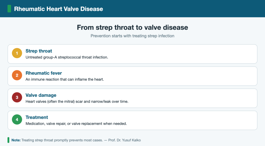
How it develops
Streptococcal bacteria carry surface components that resemble proteins in heart valve tissue. When the immune system attacks the bacteria, it can attack the valve alongside them by mistake. The episode of rheumatic fever itself passes — migrating joint pain, fever, fatigue — but the inflammation leaves scar tissue in the valve leaflets, which slowly thickens and calcifies over years. This is why a question about “repeated sore throats or joint pains in childhood” forms part of assessing an adult with unexplained breathlessness.
And every further attack adds more damage. Preventing the next one is therefore not a side note about hygiene; it is part of treating the disease itself.
Which valve, and why it matters
The mitral valve is affected far more often than any other, followed by the aortic; both can be involved together. The damage takes one of two forms: stenosis, where the leaflets fuse and the opening narrows so blood is held back behind the valve, or regurgitation, where the leaflets no longer close and blood flows backwards. Mitral stenosis is the picture most characteristic of this disease.
Symptoms
- Breathlessness on exertion, worsening gradually, and later when lying flat,
- Fatigue and reduced exercise capacity,
- Palpitation — which may be the first appearance of atrial fibrillation,
- Coughing blood-streaked sputum in advanced mitral stenosis,
- Swelling of the legs in later stages,
Pregnancy can unmask it: blood volume and heart rate both rise in pregnancy, so a mitral stenosis that had been tolerated for years begins to cause clear breathlessness in the second or third trimester. For this reason a woman with known valve disease should ideally be assessed before planning a pregnancy rather than during one.
Emergencies: sudden weakness or numbness on one side, or slurred speech (possible stroke from a clot); sudden severe breathlessness or pink frothy sputum (acute pulmonary congestion); persistent fever with sweats and fatigue in someone with known valve disease (possible infection of the valve). None of these should wait for a scheduled appointment.
Atrial fibrillation and the risk of clot
This is the most important practical complication of mitral stenosis. Blood held back behind the narrowed valve enlarges the left atrium, its rhythm becomes disorganised and atrial fibrillation appears. A clot can then form inside the atrium and travel to the brain — a recognised cause of stroke in people who are relatively young and have no other risk factors.
Anticoagulation is therefore a central part of management where atrial fibrillation or a previous clot is present, and it is not stopped on one’s own initiative. What distinguishes this situation is that the risk originates in the heart, not in the neck arteries, so it is not excluded by a carotid scan — the same point approached from the other side in carotid artery disease and arrhythmia.
Diagnosis
- Echocardiography is the basis: it assesses each valve, the degree of narrowing or leakage, the size of the atrium and the function of the heart muscle,
- ECG, to detect atrial fibrillation,
- Transoesophageal echocardiography before any intervention — specifically to exclude a clot in the atrium, because its presence rules out balloon dilatation,
- History — repeated throat infections, or an episode of fever with joint pains in childhood.
Treatment options
| Option | When it is considered | What goes with it |
|---|---|---|
| Medication | Symptom and rate control, with anticoagulation where indicated | Does not repair the valve, but controls complications and can postpone intervention |
| Balloon mitral valvuloplasty | Mitral stenosis with suitable anatomy, no significant regurgitation and no clot in the atrium | Performed by catheter without opening the chest; suitability is settled on transoesophageal echocardiography |
| Valve repair | Leaflets still suitable for reconstruction | Preserves the patient’s own valve where it is feasible |
| Valve replacement | Severe damage beyond repair | The choice of valve type balances durability against the need for lifelong anticoagulation |
One point matters particularly for women of childbearing age: the type of valve chosen affects future pregnancy planning, because some types require permanent anticoagulation, which needs special management during pregnancy. That conversation belongs before the operation, not after it.
Secondary prevention: the part that gets forgotten
Once rheumatic fever has been established, a long-acting preventive antibiotic is usually prescribed at regular intervals for years, to prevent a new streptococcal throat infection triggering another attack. How long it continues depends on age, on whether the valve was actually damaged and on how severely. Many patients stop once they feel well — and that is precisely what leads to further damage years later.
- Treat strep throat in children completely, and do not stop the antibiotic when the pain goes,
- Keep to the preventive doses on schedule and do not discontinue them on your own,
- Tell your dentist about valve disease or a prosthetic valve before any procedure,
- Look after your mouth — dental health is among the most effective ways of reducing the risk of valve infection,
- Attend follow-up echocardiography even without symptoms; narrowing progresses slowly and silently.
Why Choose Turkey (Istanbul)?
- Over 30 years of experience in cardiovascular and vascular surgery with Prof. Dr. Yusuf Kalko.
- Modern minimally invasive and hybrid techniques.
- English- and Arabic-speaking support for international patients.
- Easy access to Istanbul from the Gulf, Europe and beyond.
- Remote assessment before travel, then treatment and appointment planning.
How to Start From Abroad (Remote Consultation)
You can send your medical reports and scan images via WhatsApp for an initial assessment before traveling. We then plan your treatment and appointment in Istanbul, Turkey, and support international patients at every step.
To evaluate your heart valves in Istanbul (Beylikdüzü) and plan suitable treatment, contact us on WhatsApp.
Related Pages
Frequently Asked Questions
Is rheumatic heart disease preventable?
Largely yes — prompt treatment of strep throat prevents most cases, which is why early infection care matters.
Which valve is most affected?
The mitral valve is most commonly affected, followed by the aortic valve.
Is surgery always needed?
No. Mild disease is monitored and managed medically; surgery (repair or replacement) is reserved for significant valve damage.
Do you treat international patients?
Yes. We welcome international and Gulf patients in Turkey (Istanbul), with English- and Arabic-speaking support throughout assessment and treatment.
How can I book an appointment from abroad?
Via WhatsApp: send your inquiry and medical reports, and we will arrange your appointment and explain the steps before and after arriving in Istanbul.
Can I have a consultation before traveling?
Yes. An initial remote assessment can be made based on your reports and scans before you come to Turkey.